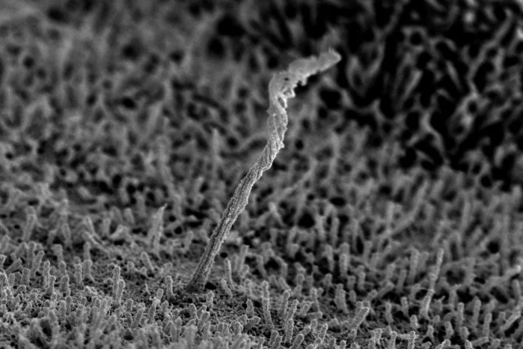
Three dimensional images of human pancreatic islet cells provide an unprecedented view of the enigmatic primary cilia

Jing Hughes, MD, Assistant Professor of Medicine, Division of Endocrinology, Metabolism & Lipid Research recent paper, “Scanning electron microscopy of human islet cilia” was published in Proceedings of the National Academy of Sciences (PNAS) and follow-up article, “Immuno-scanning electron microscopy of islet primary cilia,” in the Journal of Cell Science.
The Hughes Lab PNAS publication was also cited by The Scientist in “Science Snapshot,” which details the discovery of a 3D structure of the primary cilium of the human islet, which was completed in collaboration with the Department of Cell Biology & Physiology and the Center for Cellular Imaging at Washington University.
Additionally, Dr. Hughes was also interviewed by The Scientist regarding her work studying the structure of the cilia on the surface of the pancreas. Hughes hopes to understand their basic functions, which may help scientists determine how cilia defects can lead to disease. “It’s a peculiar structure. It’s long, it’s awkward, it’s vulnerable to a lot of mechanical stress, but certainly it has evolved to be that way to perfect a function that it serves,” Hughes said. “It really captured my imagination.”
